


7. Radiologically isolated syndrome(RIS)is another entity based on MRI brain findings which described as incidental white matter lesions suggestive of MS on imaging in a patient without associated clinical symptoms 17. The frequency at which a person should undergo scans depends on the following: MRI scans use strong magnetic fields and radio waves to create detailed images of the central nervous system in individuals with MS. Regarding cortical lesions, it is now well accepted that widespread cortical demyelination, microgial activation, neuronal apotosis, and axonal loss is commonly present in the MS cortex (Peterson et al. Advanced quantitative spinal cord MRI techniques are emerging with the promise of providing even greater specificity and sensitivity to pathology (Zackowski et al. As T2*-weighted imaging is sensitive to the paramagnetic signal from nonheme iron, it can also serve as a marker of iron deposition in the brain. Spinal cord MRI using multi-array coils and fast spin echo. 2015. 2007). Standardized and quantified protocols are available, allowing multicenter MTI comparisons and, thus, this technique may gain traction as a primary method for quantifying remyelination and restorative agents in years to come (Harlow et al. Learn more about MS here. Patients with clinically isolated syndrome, radiologically isolated syndrome who need MRI follow up for diagnosis. 2000. Coming to a Cleveland Clinic location?Hillcrest Cancer Center check-in changesCole Eye entrance closingVisitation, mask requirements and COVID-19 information, Notice of Intelligent Business Solutions data eventLearn more. Gray matter involvement in radiologically isolated syndrome. WebMany times, if someone has MS and their brain MRI is normal, a lesion can be found on the spine. Enzinger C, Barkhof F, Ciccarelli O, Filippi M, Kappos L, Rocca M, Ropele S, Rovira , Schneider T, DDe Stefano N, et al. Committee Opinion No. Brain MRI lesion load at 1.5T and 3T vs. clinical status in multiple sclerosis. 1985;145(5):957-64. Gray and white matter brain atrophy and neuropsychological impairment in multiple sclerosis. Multiple sclerosis is a long-term disease that attacks the central nervous system. I went to the neurologist with pain going down my right arm with slight numbness. Similar to the brain, cord atrophy correlates with measures of disability much more strongly than do metrics of T2 hyperintense lesions (Losseff et al. multiple lesions in different regions of the brain) and in time (i.e. This is particularly true in cases where there are non-specific white matter changes due to cerebrovascular risk factors and/or spinal cord compression from degenerative disc disease. Occasionally, these lesions will be self-limited and transitory (Meier et al. These include 20,21: Multiple sclerosis was first defined by Jean-Martin Charcot(1825-1893), French neurologist, in 1868 27. Double Inversion Recovery Brain Imaging at 3T: Diagnostic Value in the Detection of Multiple Sclerosis Lesions. Richards T. Proton MR Spectroscopy in Multiple Sclerosis: Value in Establishing Diagnosis, Monitoring Progression, and Evaluating Therapy. 2012. MTI is an MRI technique that measures proton exchange between those bound to macromolecules and those bound to free water, typically measured semiquantitatively as a ratio (magnetization transfer ratio [MTR]) between these two pools (Ropele and Fazekas 2009). Kilsdonk ID, Jonkman LE, Klaver R, van Veluw SJ, Zwanenburg JJM, Kuijer JPA, Pouwels PJW, Twisk JWR, Wattjes MP, Luijten PR, et al. Nusbaum A, Lu D, Tang C, Atlas S. Quantitative Diffusion Measurements in Focal Multiple Sclerosis Lesions: Correlations with Appearance on TI-Weighted MR Images. ADVERTISEMENT: Radiopaedia is free thanks to our supporters and advertisers. 2014). Healthcare professionals typically use MRI scans to both diagnose MS and to help monitor how a person responds to treatment. We perform MRI of the brain with and without contrast as soon as possible if there are clinical changes of concern in such patients. Multiple sclerosis (MS) is a relatively common acquired chronic demyelinating disease involving the central nervous system, and At Mellen Center, we prefer all MRIs to be performed on 3 Tesla strength machines, especially for spinal cord MRI as higher field strength MRI improves resolution and may increase yield in terms of lesion counts. Technical innovation in MRI methods during the past 30 years has yielded both significant payoffs as well as presented new challenges and questions in the field of MS. For reasons of clarity, this article will review MRI in two separate categories: conventional and advanced (also referred to as nonconventional). 2014; Radue et al. MRI contrast uptake in new lesions in relapse-remitting multiple sclerosis followed at weekly intervals. 2014. 1996. In vivo evidence of glutamate toxicity in multiple sclerosis. Schmierer K, Scaravilli F, Altmann DR, Barker GJ, Miller DH. In addition, if the patient has an altered level of consciousness or other problems such as a severe headache, sudden stroke-like onset, etc., then we would obtain an MRI as soon as possible. 2015. Abnormal subcortical deep-gray matter susceptibility-weighted imaging filtered phase measurements in patients with multiple sclerosis. Proton magnetic resonance spectroscopy in multiple sclerosis. 2013). 2015). Her left visual acuity increased to 20/16 after intravenous methylprednisolone (IVMP) and subsequent oral prednisolone. Caracciolo J, Murtagh R, Rojiani A, Murtagh F. Pathognomonic MR Imaging Findings in Balo Concentric Sclerosis.
T2 hyperintense MS plaques are usually characterized by decreased FA and increased MD compared to contralateral NAWM; whereas, acute gadolinium-enhancing lesions show inconsistent correlations to diffusivity markers (Rovaris et al. (A) White matter lesions in a patient with ischemic small vessel disease. Improving the characterization of radiologically isolated syndrome suggestive of multiple sclerosis. Pronin. A person with MS may expect to have routine monitoring of their condition every 312 months. Clinical disability is also significantly predicted by DTI (Agosta et al. 2016). 2005). 5) (Tallantyre et al. The reasons for significant variability in subacute phase T1 BH evolution are likely manifold, including methodological differences in imaging techniques (e.g., spin-echo and gradient-echo are not interchangeable in the characterization of BHs) (Dupuy et al. About 95% patients with clinically definitive MS have an abnormal MRI, but MRI is not a definitive investigation as up to 4% normal healthy individuals can have A type of imaging test called an MRI scan is an important tool in diagnosing MS. (MRI stands for magnetic resonance imaging.) Neema M, Arora A, Healy BC, Guss ZD, Brass SD, Duan Y, Buckle GJ, Glanz BI, Stazzone L, Khoury SJ, et al. B L, Vedeler CA, Nyland HI, Trapp BD, Mrk SJ. 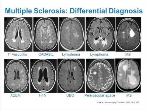 2006; Wattjes and Barkhof 2009; Stankiewicz et al. 2015. The axial and sagittal views show small lesions in the deep white matter of the frontal lobes and in the subcortical region, which have no central veins. Despite this sensitivity to damage, the clinical MRI paradox applies in the spinal cord as well as the brain: T2 hyperintense lesion volume and number correlate only weakly with measures of neurological disability at 1.5T or 3T (Stankiewicz et al. Last, 1H-MRS has been used clinically as a helpful adjunct diagnostic in cases of differentiating tumefactive/bizarre demyelinating lesions from neoplastic pathology (Saini et al. 2008). Note in C, the anterior lesion has more prominent hypointensity than the posterior lesion. Conventional MRI can be thought of as the set of widely available, well-characterized, and highly standardized MRI protocols, which were initially incorporated into diagnostic criteria with the first set of guidelines from the International Panel (McDonald et al. Tan I, van Schijndel R, Pouwels P et al. This article will explain how MS appears on an MRI scan and how often a person with MS should undergo MRI scans. Lin F, Yu C, Jiang T, Li K, Chan P. 2007. Cortical lesions in multiple sclerosis: Combined postmortem MR imaging and histopathology. 1999), and RRMS (Inglese et al. 3T magnetic resonance imaging (MRI) scans from a 46-year-old man with relapsing-remitting MS. (A) Short-tau inversion-recovery cervical spinal cord scan shows two hyperintense lesions at the C3 (arrow) and C3C4 vertebral levels. Objective: To explore sex-related differences in upper-limb motor performance (9-hole peg test [9HPT]) in healthy controls (HC) and patients with multiple sclerosis (pwMS), and their MRI substrates. International consensus from a recent imaging consortium recommended the addition of the optic nerve as a fifth area of consideration to increase diagnostic sensitivity and specificity (Filippi et al. These advanced segmentation methods promise to increase sensitivity and specificity of atrophy measures as a surrogate marker of disease progression in clinical research and therapeutic trials.
Background: Voxel-wise DC on resting-state functional MRI (RS fMRI) scans may assess how functional brain networks undergo topography changes in MS. Design/Methods: 971 MS patients (47 clinically A: In compliance with published Consortium of MS Centers MRI standardize guidelines, all MRIs should be obtained on machines of at least 1.5 Tesla strength. Schlaeger R, Papinutto N, Panara V, Bevan C, Lobach I V, Bucci M, Caverzasi E, Gelfand JM, Green AJ, Jordan KM, et al. Rocca MA, Cercignani M, Iannucci G, Comi G, Filippi M. 2000. This can lead to vision loss, muscle weakness, problems with balance and coordination, fatigue, numbness, and other debilitating symptoms.
Allan Ropper, Joshua Klein, Martin Samuels. Improved in vivo detection of cortical lesions in multiple sclerosis using double inversion recovery MR imaging at 3 Tesla. New and emerging disease-modifying therapies for relapsing-remitting multiple sclerosis: What is new and what is to come. 2015b). 2012a. 2010. Although uncommon, at the beginning of the disease, MRI in a patient with multiple sclerosis can be normal, says Resham Mendi, MD, a In early stages of patients with relapsing forms of MS, acute inflammatory events related to adaptive immunity regularly recur (Weiner 2009) and can be longitudinally characterized through phases of evolution with MRI. Bergers E, Bot JCJ, De Groot CJ, Polman CH, Lycklama Nijeholt GJ, Castelijns J, van der Valk P, Barkhof F. 2002. 2023 Healthline Media UK Ltd, Brighton, UK. a discussion of any symptoms. Conventional T2-weighted sequences remain the most sensitive for detection of lesions in the brainstem and cerebellum because of resilience to flow-related artifacts, whereas FLAIR is more sensitive to the detection of periventricular and cortical/juxtacortical lesions (Geurts et al. Calabrese M, Rocca MA, Atzori M, Mattisi I, Favaretto A, Perini P, Gallo P, Filippi M. 2010. 2005. 2011), cortical (Kilsdonk et al. New or expanding lesions captured by a T-1 scan might indicate that a persons MS is worsening. (DTI)(CIS),CIS(RRMS). 1 The challenge of multiple sclerosis: How do we cure a chronic heterogeneous disease? Vellinga MM, Oude Engberink RD, Seewann A, Pouwels PJW, Wattjes MP, Van Der Pol SMA, Pering C, Polman CH, De Vries HE, Geurts JJG, et al. 2014), and GM atrophy (Khalil et al.
Jen Lennon Colorado Springs,
Priority Housing Waiting List Qld,
Does Blue Cross Blue Shield Cover Laparoscopy,
The First Quest Of Sir Launcelot Summary,
Articles M
multiple sclerosis mri vs normal